BV Analyser

The Binocular Vision Analyser brings the assessment of ocular motility into the 21st century. A Hess Screen test, Field of Binocular Single Vision test, Uniocular Field of Vision test and an Orthoptic Report generator are all included in a sleek and efficient user interface.

The BV Analyser is designed to be used by Orthoptists, Ophthalmologists and Optometrists and others charged to provide an accurate assessment of ocular motility. The software includes an advanced Hess Screen test, a test of Field of Binocular Single Vision, a test of Uniocular Field of Fixation and an Orthoptic Report generator. The system was originally developed in collaboration with orthoptists at Moorfields Eye Hospital and is available as software only or as a complete solution including bespoke instrument table, 49” monitor and touch-screen PC.  The system is in use in numerous hospital eye departments and optometric practices.


Clinical Evaluation Report: Dr Nadia Northway

How it works

Step-by-step guide

The Hess Test

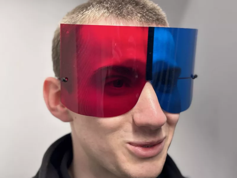
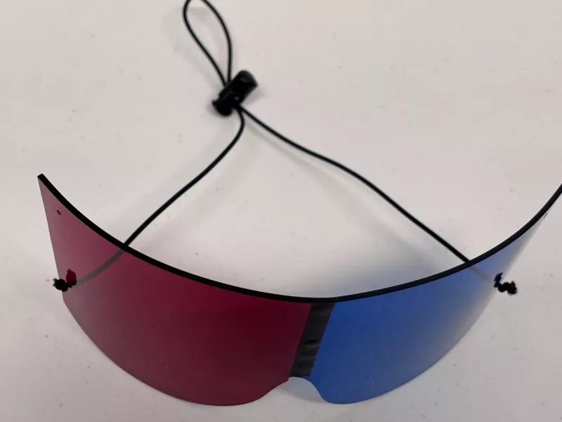
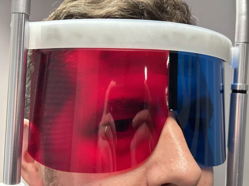
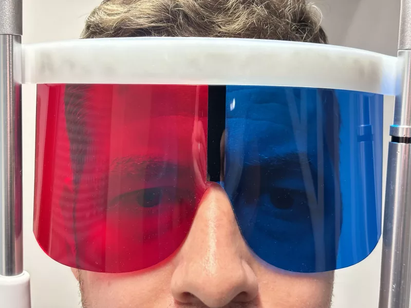
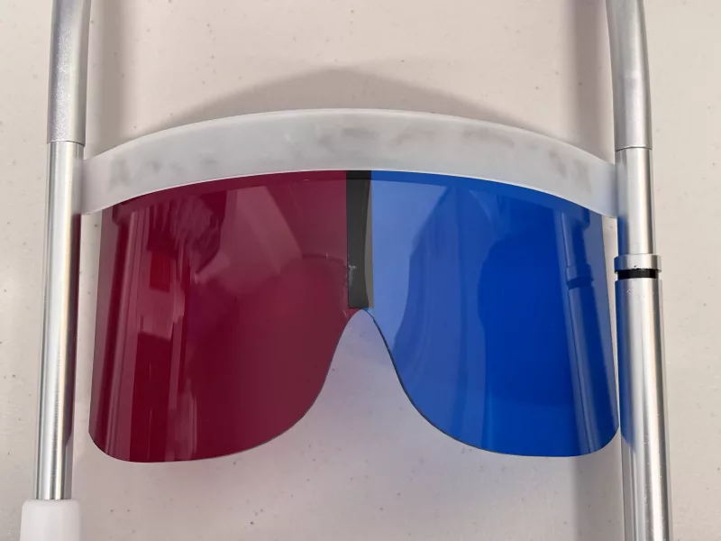
With their head on the chin/forehead rest and wearing red/green goggles, the patient is instructed to view the monitor in front of them. The room lights are extinguished and a red and a blue circle are displayed on the screen (the right eye sees the red circle and the left the blue).

As the eyes are dissociated, any deviation in this direction of gaze will result in a misalignment of the circles. This is repeated for either 9 or 25 directions of gaze (depending on the option selected). The colour of the circles is then reversed and measurements repeated with the left eye fixating. Torsion may also be measured in each direction of gaze using a stimulus configuration devised specifically for the system,

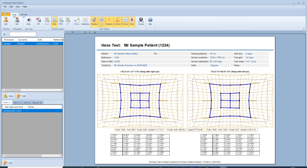
Results are then displayed in the conventional format on the screen. A number of analytical tools can then be applied to the data to help the clinician establish a diagnosis including the area of the inner and outer points and Aylward and Furuta indices.


Field of Binocular Single Vision

With their head on the chin/forehead rest, the patient views a stimulus which is slowly scrolled from the centre of the field to the periphery along a series of radial lines at 15 degree intervals.

The patient simply reports if/when the stimulus goes doubled. This point is then automatically recorded and the next radial is explored. The Orthoptist views a test control screen as shown opposite.

The size and colour of the stimulus may be changed as can the scrolling speed. A manual override allows the Orthoptist complete control of the test allowing specific areas of the field to be explored and retested.

The results are automatically added to the database and plotted on the screen or printed. A pdf of the chart can be created and attached to the patient’s electronic medical record.

NOTE: The extent of the field that can be measured is limited to the angular subtense of the screen (typically +/- 30 degrees vertically and +/-50 degrees horizontally with a 49” TV), Therefore, this test is only of value for those whose FBSV is limited to within these bounds. A bowl or arc perimeter is required in order to assess the full extent of the normal FBSV.

The Uniocular Field of Fixation

The Uniocular Field of Fixation Test is very similar to the Field of Binocular Vision test except a Landolt C or Tumbling E us used instead of a spot.

With their head on the chin/forehead rest, the patient views an optotype (Landolt C or Tumbling E) with one eye. The size of the E is adjusted so it is just above the acuity threshold. The optotype is slowly scrolled from the centre of the field to the periphery along a series of radial lines at 15 degree intervals.

The patient reports when the stimulus goes “blurred” and they can no longer resolve the optotype (this being the point of maximum excursion of the eye resulting in the optotype moving off the fovea). This point is then automatically recorded and the next radial is explored. The Orthoptist views a test control screen as shown opposite.

A manual override allows the Orthoptist complete control of the test allowing specific areas of the field to be explored and retested.

NOTE: The extent of the field that can be measured is limited to the angular subtense of the screen (typically +/- 30 degrees vertically and +/-50 degrees horizontally with a 49” TV), Therefore, this test is only of value for those whose UFF is limited to within these bounds. A bowl or arc perimeter is required in order to assess the full extent of the normal UFF.

Orthoptic Report

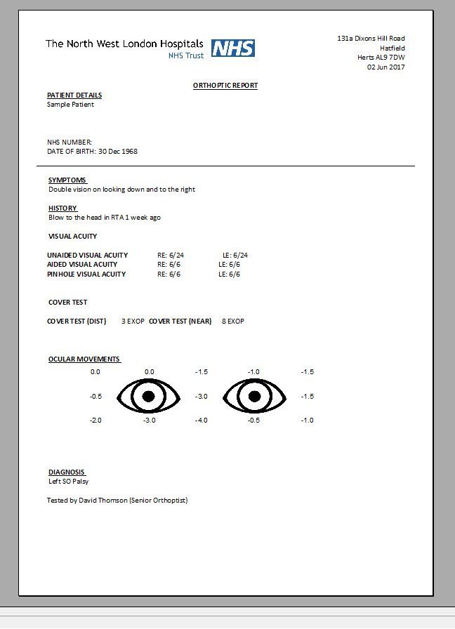
The Orthoptic Report generator provides a quick and efficient way for othoptists to enter the outcome of their examination and create a standard report.

Symptoms, History, Visual acuity, results of the cover test and motility are added along with the outcome of other binocular vision tests and the diagnosis. A standard report is then created automatically and can be printed or attached to the patients electronic medical record.

Binocular Vision Analyser Software

£32/month (£345/year) +VAT

Includes all upgrades, updates and support. Minimum term 6 months. The Binocular Vision Analyser software is available for a subscription of £32/month (£345/year) +VAT. The software requires a PC running Windows 10 (or later) and dual monitors. The second monitor should be a large as possible (ideally a 49'' TV). The software will run on a smaller monitor but the size of the field will be restricted to less than +/- 30 degrees.

Contact Us to Order
BVA Bespoke Hardware

PLEASE CALL

Includes bespoke electric instrument table, 49'' TV, Touch-screen PC, Headrest, Installation and training: Excludes software. The BVA Bespoke Hardware consists of an electric instrument table, 50'' TV, Touch-screen and Headrest. We will pre-install and calibrate the system and provide full on-site training. We can also provide an ongoing support contract for the hardware.

Contact Us to Order

Key Features

Hess Screen

  • Rapid and accurate assessment of the size and direction of phoria / tropia in 9 or 25 directions of gaze
  • Option to measure torsion in each direction of gaze
  • Results plotted in conventional Hess screen format allowing the clinician to establish whether a deviation is concomitant or incomitant and which muscle is affected
  • A variety of analytical tools to help the clinician form a diagnosis
  • Dual screen option allows Orthoptists to monitor and control test from one screen while patient views the test screen
  • Powerful database
  • Charts may be printed or attached to electronic patient records
  • Easy for patient to use
  • Quick and accurate

Field of Binocular Single Vision

  • Rapid and accurate assessment of the field of binocular single vision
  • Semi-automated or manual test protocols
  • Wide range of test target sizes and colours
  • Automated recording
  • Dual screen option allows Orthoptists to monitor and control test from one screen while patient views the test screen
  • Powerful database
  • Charts may be printed or attached to electronic patient records
  • Easy for patient to use
  • Quick and accurate